Share :
Atopic dermatitis (AD) is a chronic inflammatory skin disease affecting up to 20% of children and 10% of adults worldwide. It is characterized by itching, inflammatory lesions, and dry skin. A key factor in this condition is excessive colonization of the skin by Staphylococcus aureus, which disrupts the skin barrier and exacerbates inflammation. This colonization is frequently observed in lesions and even on unaffected skin areas. At the same time, skin microbial diversity is often reduced, limiting the presence of beneficial commensal microbes (1–5).
AD results from a complex interaction between genetic predispositions, alterations of the skin barrier, dysbiosis of the skin microbiome, and environmental factors (3,6). Barrier dysfunction is linked to genetic mutations, notably in the FLG gene encoding filaggrin, which is essential for skin hydration and protection. This mutation, in addition to downregulating certain structural proteins, promotes antigen sensitization (6). AD is also associated with an imbalanced immune response dominated by Th2 and Th17 cytokines (3). Epigenetics and the exposome, including the influence of environmental pollutants and oxidative stress, play a crucial role in the disease’s pathogenesis. The “atopic march,” a phenomenon encompassing the transition from atopic dermatitis to other allergic conditions such as asthma and rhinitis, illustrates how atopic symptoms can evolve from a very young age (1).
Beyond Staphylococcus aureus, other microorganisms influence the course of atopic dermatitis. Staphylococcus epidermidis usually plays a protective role by limiting the proliferation of S. aureus through the production of antimicrobial molecules; however, its presence is often reduced in dysbiosis. Byrd et al. showed that milder AD flares were associated with higher levels of S. epidermidis, whereas more severe flares correlated with S. aureus. Conversely, certain yeasts of the genus Malassezia may contribute to worsening symptoms, particularly during inflammatory flares (6,7,8).
Management of AD relies on conventional therapies such as topical corticosteroids and antibiotics, as well as practices like diluted bleach baths (9). Innovative approaches include autologous microbiotherapy, the use of probiotics and prebiotics to rebalance the skin microbiome, and JAK inhibitors to reduce inflammation (6). A better understanding of these factors will enable the development of personalized treatments aimed at improving patients’ quality of life. Microbiome-friendly treatments may also be very beneficial for atopic skin. Strategies that reduce S. aureus colonization while promoting the expansion of S. epidermidis or other beneficial commensal bacteria also represent a promising avenue for new treatments.
At the request of a client developing a cream for atopic-prone skin, BYOME LABS designed a bespoke protocol to evaluate, through in vitro tests, its impact on Staphylococcus aureus in both planktonic and biofilm forms. Our experts selected the relevant strains based on the scientific literature and data from other microbiome study techniques (NGS sequencing). After an initial phase of planktonic testing, the study focused on the cream’s preventive effect on biofilm formation. Result: the product did not show a bactericidal effect, but it demonstrated an ability to limit biofilm formation, making S. aureus colonization more transient.
At BYOME LABS, our teams tailor their know-how to your products and objectives and support you throughout the entire process.
Sources :
1. Spergel JM, Paller AS. Atopic dermatitis and the atopic march. J Allergy Clin Immunol. 1 déc 2003;112(6):S118‑27.
2. Centre de Preuves de la Société Française de Dermatologie [Internet]. [cité 9 avr 2025]. Disponible sur: https://centredepreuves.sfdermato.org/
3. Kobayashi T, Glatz M, Horiuchi K, Kawasaki H, Akiyama H, Kaplan DH, et al. Dysbiosis and Staphylococcus aureus colonization drives inflammation in atopic dermatitis. Immunity. 21 avr 2015;42(4):756‑66.
4. Kong HH, Oh J, Deming C, Conlan S, Grice EA, Beatson MA, et al. Temporal shifts in the skin microbiome associated with disease flares and treatment in children with atopic dermatitis. Genome Res. mai 2012;22(5):850‑9.
5. Grice EA, Kong HH, Conlan S, Deming CB, Davis J, Young AC, et al. Topographical and Temporal Diversity of the Human Skin Microbiome. Science. 29 mai 2009;324(5931):1190‑2.
6. Savva M, Papadopoulos NG, Gregoriou S, Katsarou S, Papapostolou N, Makris M, et al. Recent Advancements in the Atopic Dermatitis Mechanism. Front Biosci-Landmark. 22 févr 2024;29(2):84.
7. Song YC, Hahn HJ, Kim JY, Ko JH, Lee YW, Choe YB, et al. Epidemiologic Study of Malassezia Yeasts in Acne Patients by Analysis of 26S rDNA PCR-RFLP. Ann Dermatol. août 2011;23(3):321‑8.
8. Byrd A. L. et al. (2017). Staphylococcus aureus and Staphylococcus epidermidis strain diversity underlying pediatric atopic dermatitis. Science translational medicine.
9. Kantor R, Silverberg JI. Environmental risk factors and their role in the management of atopic dermatitis. Expert Rev Clin Immunol. janv 2017;13(1):15‑26.
Did this article appeal to you and would you like to find out more about this topic?
This site is protected by reCAPTCHA and the Google Privacy Policy
and Terms of Service apply.
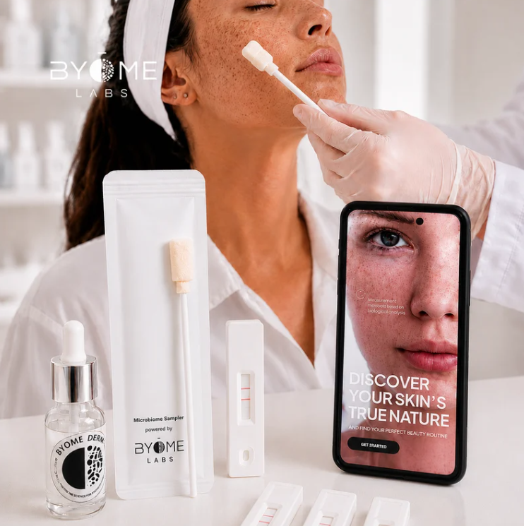
With Byome Derma, our goal is to make microbiome analysis a routine tool for better understanding individual skin needs.
In this article, we explore the brain gut skin axis and its implications for the understanding and treatment of skin disorders.

An article that looks back at our mission: supporting brands in evaluating the impact of their products on human microbiomes, and sharing our outlook around BYOME DERMA.
Notre site utilise des cookies pour améliorer votre expérience de navigation. En continuant à utiliser notre site, vous acceptez notre utilisation de cookies conformément à notre politique de confidentialité.